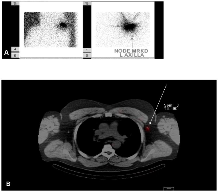
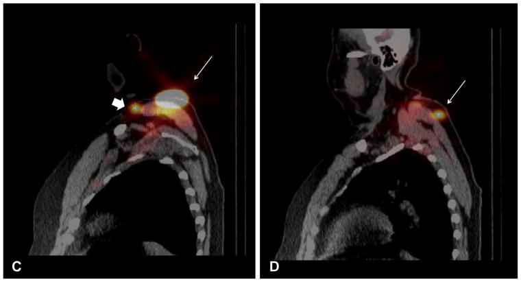
FIG. 2.
Planar lymphoscintigraphy revealed a left posterior shoulder primary with one node marked in the left axilla (A). SPECT/CT demonstrated one node located in the left axilla (white arrow) (B). SPECT/CT also demonstrated the left shoulder primary (white arrow) along with a left supraclavicular node (arrow head) (C) and a left posterior superficial subcutaneous node (white arrow) (D).


