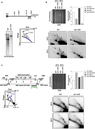
Fig. 4. Stn1 is required to promote efficient replication of telomeric regions.

(A) Relative position of the restriction sites in the subtelomeric regions of chromosomes I and II. The subtelomeric probe (STE1) that is used for 2D-gel hybridization (see below) is represented. Cen, centromere. Southern blot analysis of Nsi I telomeric fragments (first dimension) from the parental WT strain and stn1-226 mutant revealed by STE1 probe. (B) 2D-gel analysis of Nsi I telomeric fragments of WT and stn1-226 strains at 25°C and after 24 hours at 36°C. The first dimension of the EtBr-stained gel was photographed before the second dimension. The Y-arc pattern is generated by unidirectional movement of a replication fork across each telomeric fragment shown in the first dimension. The cone-shaped signal represents four-way DNA junctions (double Y). Quantification of the total replication intermediate signal over the linear arc signal of rDNA is presented. N.A., not available. (C) Top: Map of the rDNA repeats. Boxes indicate the ars3001 and the RFB pause sites (P). The restriction enzyme sites are indicated (H, Hind III; B, Bam HI; K, Kpn I; S, Sac I; E, Eco RI). Left: Diagram of the migration pattern of replication intermediates that can be detected by 2D-gel electrophoresis. Right: 2D-gel analysis of rDNA RFB site in WT and stn1-226 strains at permissive and restrictive temperatures. The first dimension of the EtBr-stained gel was photographed before the second dimension. Quantification of the total replication intermediate signal over the linear arc signal is presented. The Eco RI–Eco RI fragment is used as a probe.
