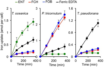
Fig. 1. Iron uptake from different iron sources by T. oceanica, P. tricornutum, and T. pseudonana.

The figure shows specificity of siderophore uptake in three species of diatoms. The cells were precultured in iron-deficient medium, harvested at mid-exponential growth, and washed once with iron-free medium. The cells were then resuspended in growth medium containing one of the following 55Fe-labeled sources supplied at 1 μM: ferric EDTA (black), FCH (red), FOB (blue), or ENT (green). The cells were washed on filters at intervals with the washing buffer, and 55Fe associated to the washed cells was counted by scintillation. Data are means ± SD from four experiments (biological replicates).
