
Figure 3. OAS treatment affects neointimal morphology.
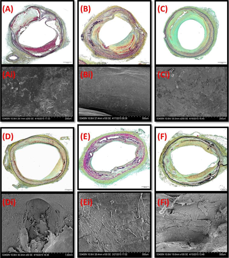
Representative MP stained cross sections (A-F) and matching enface SEM (Ai-Fi) of untreated (A-C) and OAS-treated (D-Fi) artery segments. (A,D) the SFA, (B,E) popliteal arteries, (C,F) tibial arteries.