
Figure 3.
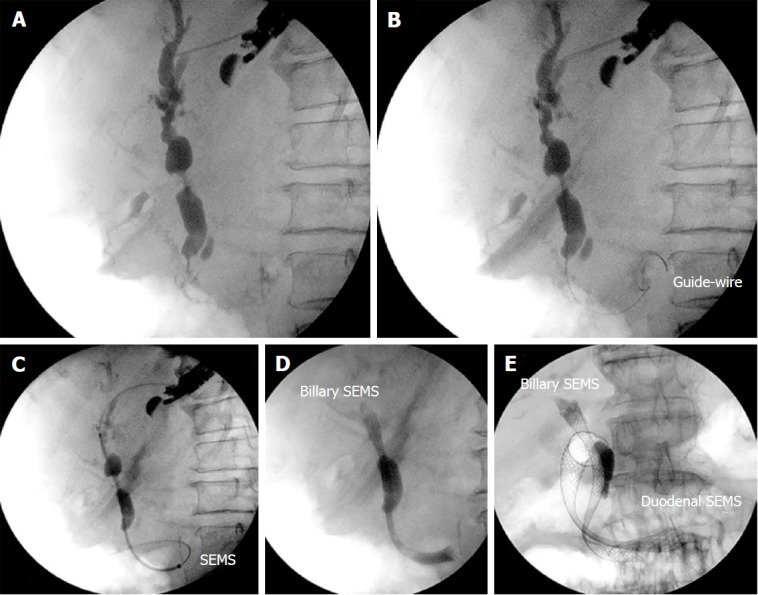
Patient with duodenal stenosis due to a pancreatic carcinoma. A: Endosonography (EUS)-guided cholangiography; B: Insertion of the guidewire through the duodenal major papilla and positioning in the duodenum; C: Anterograde insertion of the self-expandable metallic stents (SEMS) through the gastric wall across the duodenal major papilla and its positioning in the duodenum; D: Deployment of the SEMS; E: Insertion of the duodenal SEMS. SEMS: Self-expandable metallic stents.