Fig. 3.
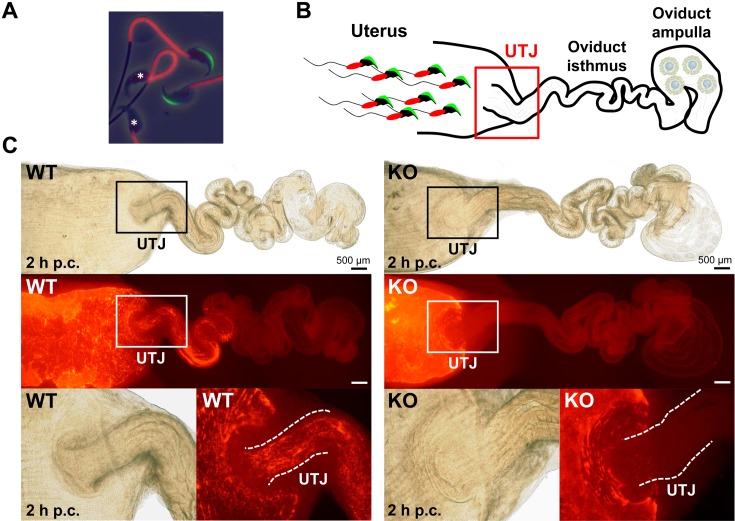
Observation of ejaculated spermatozoa into the female reproductive tract. (A) Visualization of the acrosome and midpiece of spermatozoa. A transgenic mouse line carrying Acr-Egfp and CAG-Su9/DsRed2 transgenes expressed both a green sperm acrosome and red mitochondria in the sperm midpiece [31]. It is easy to determine if the acrosome reaction occurred in these spermatozoa due to the green acrosome. *Acrosome-reacted spermatozoa. This transgenic mouse line [B6D2-Tg (CAG/su9-DsRed2, Acr3-Egfp) RBGS002Osb] is available from the RIKEN BioResource Center and the Center for Animal Resources and Development (CARD), Kumamoto University. (B) Scheme of observing sperm migration into the female reproductive tract using fluorescent spermatozoa. (C) Observation and visualization of ejaculated spermatozoa into the female reproductive tract two hours post coitus (p.c.). Observing the red signals, wild-type (WT) spermatozoa passed through the uterotubal junction (UTJ), but Tex101 KO spermatozoa were unable to migrate from the uterus to the oviduct [29]. Fluorescent spermatozoa could facilitate live imaging of localization and movement in vitro and in vivo.

