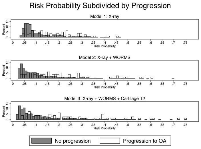
Figure 4.
The model classification improves from Model 1 (radiography + Risk factors) to Model 3 (Model 1 + WORMS + T2), as shown by the increasing spread of the data. The higher the risk probability, the higher the likelihood for progression – this phenomenon is especially pronounced when comparing Models 1 to Model 3.

