
Figure 3.
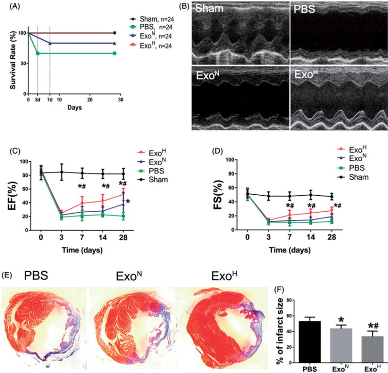
Augmenting cardiac function and ameliorating fibrosis after MI by ExoH. (A) The number of survival animals was recorded for each day of the study period (from 0 to 28 d). (B) Representative echocardiography images showing significantly increased wall motion in ExoH treated hearts. (C,D) Gradually increased ejection fraction (EF) and fractional shortening (FS) in mice transplanted with ExoH compared with other groups (n = 24 for Sham, 16 for PBS, 20 for ExoN and 24 for ExoH). (E) The cross-sections of infarcted hearts were analysed with Masson trichrome staining at 4 weeks after infarction. The fibrosis in the scar of infarcted hearts was shown in blue. (F) The fibrotic scar areas were quantified (n = 6). *p < .05, vs. PBS; #p < .05, vs. ExoN.