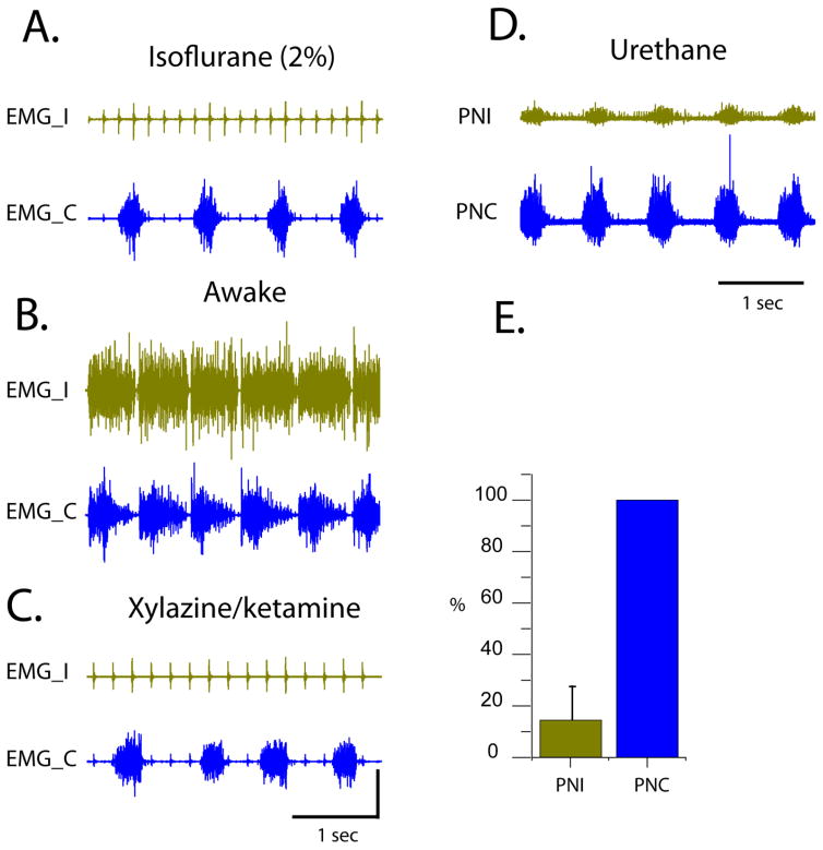
Figure 4.
A. An example of a bilateral diaphragm EMG recording in a rat under isoflurane anesthesia (8 weeks post-C2Hx); B. An example of a bilateral diaphragm EMG recording 10 min post-isoflurane anesthesia (8 weeks post-C2Hx); C. An example of a bilateral diaphragm EMG recording in a rat under xylazine/ketamine anesthesia (8 weeks post-C2Hx); D. An example of a bilateral phrenic recording in a rat under urethane anesthesia (8 weeks post-C2Hx). Panels A–D represent recordings from the same rat. E. Summary histogram of averaged phrenic integrated amplitudes from the ipsilateral (PNI) and contralateral (PNC) sides. Ipsi- amplitude is normalized to the contrlateral side. EMG_I – ipsi EMG, EMG_C – contra EMG, PNI – ipsilateral phrenic neurogram, PNC – contralateral phrenic neurogram.

