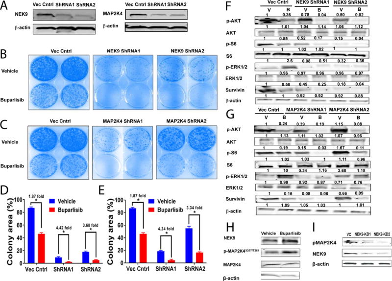
Figure 7. In vitro target validation of NEK9 and MAP2K4 in association with buparlisib resistance.

A, Western blot showing knockdown of either NEK9 or MAP2K4 in the WHIM12 cell line. B and C, colony formation assays of WHIM12 cells with NEK9 or MAP2K4 knocked down, respectively. D and E, quantified growth of colony formation assays in panel B (NEK9) and C (MAP2K4), respectively. F, Western blot of a set of relevant PI3K markers after NEK9 knockdown in the WHIM12 cell line – with or without buparlisib treatment. G, Western blot of a set of relevant markers after MAP2K4 knockdown in the WHIM12 cell line – with or without buparlisib treatment. H, NEK9 protein levels and MAP2K4 S257/T261 phosphorylation (pMAP2K4) after buparlisib treatment. I, NEK9 knockdown in the WHIM12 cell line leads to loss of MAP2K4 S257/T261 phosphorylation. V: Vehicle; B: buparlisib; VC: Vector Control; KD1: knockdown clone 1; KD2: knockdown clone 2. * p ≤ 0.05. All analyses performed on a cell line derived from the WHIM12 xenograft.
