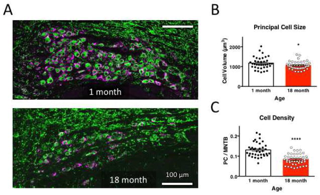
Figure 1. MNTB neuron size and density is attenuated with age.
A. Neurons in fixed coronal slices from 1-month (top) and 18-month old mice (bottom) were identified by anti-MAP2 immunoreactivity, and the presynaptic calyceal terminal stained with vGluT1. Images have shared orientation, with midline to the right, dorsal up. Scale bar is 100 μm in both images. B. Neuronal cell size was evaluated as volume of MAP2-positive signal in 9–15 μm thick confocal stacks, and is significantly reduced at 18 months, relative to neurons in 1-month old MNTB. C. Neuronal cell density was evaluated as the area occupied by MAP2-positive signal within the MNTB nucleus, and was substantially reduced in older mice.

