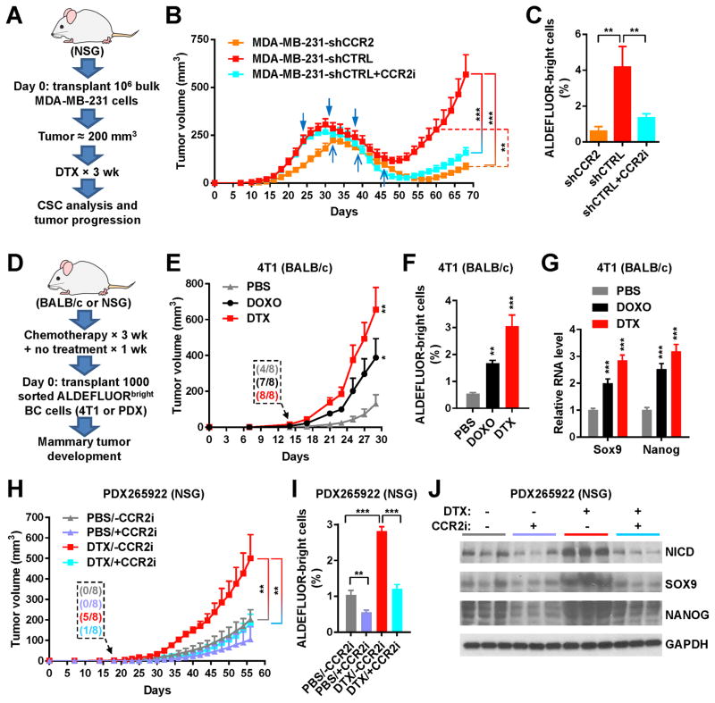
Figure 4. Chemotherapy promotes CSC properties in vivo through MCP-CCR2 signaling.
(A) Schema of the mouse model used to examine if CCR2 intervention suppresses post-chemotherapy tumor progression. One million of MDA-MB-231 cells with stable knockdown of CCR2 (shCCR2) or those expressing control shRNA (shCTRL) were injected into the #4 mammary fat pad of NSG mice. When tumor size reached ~250 mm3, mice were treated with DTX for 3 weeks, and then left free of chemotherapy until the end of experiment. One group with MDA-MB-231-shCTRL tumors also received the CCR2 inhibitor MK-0812 starting with the chemotherapy and continuing for a total of 30 days, whereas the other two groups received the vehicle. (B) Tumor onset and volume (n=8). The two groups with MDA-MB-231-shCTRL tumors received 3 times of treatment with DTX on days 24, 31, and 38 (blue arrowheads); the group with MDA-MB-231-shCCR2 tumors received DTX on days 32, 39, and 46 (blue arrows). (C) ALDEFLUOR assay of dissociated MDA-MB-231 tumor cells. (D) Schema of the mouse models used to examine if chemotherapy prior to BC cell engraftment enhances tumor formation. BALB/c and NSG mice were treated with DOXO, DTX, or PBS for 3 weeks and then left free of chemotherapy for 1 week, before 1000 FACS-isolated ALDEFLUORbright 4T1 or PDX265922 cells were injected into the #4 mammary fat pad to assess tumor development. (E) Tumor onset and volume for the 4T1 model in BALB/c mice (n=8). Inset shows numbers of mice with palpable tumors on day 14. (F) ALDEFLUOR assay of dissociated 4T1 tumor cells. (G) Relative RNA levels of indicated genes (normalized to Gapdh) in 4T1 tumor tissue determined by quantitative RT-PCR assay. (H) Tumor onset and volume for the PDX265922 model in NSG mice (n=8). CCR2 inhibitor MK-0812 or vehicle was orally administered at 30 mg/kg twice a day starting with the chemotherapy and continuing for 30 days after BC cell engraftment. Inset shows numbers of mice with palpable tumors on day 18. (I) ALDEFLUOR assay of dissociated PDX265922 tumor cells. (J) Western analysis showing indicated protein levels in PDX265922 tumor tissue. *P<0.05, **P<0.01, ***P<0.001 (compared to the corresponding PBS group in E–G or as indicated).

