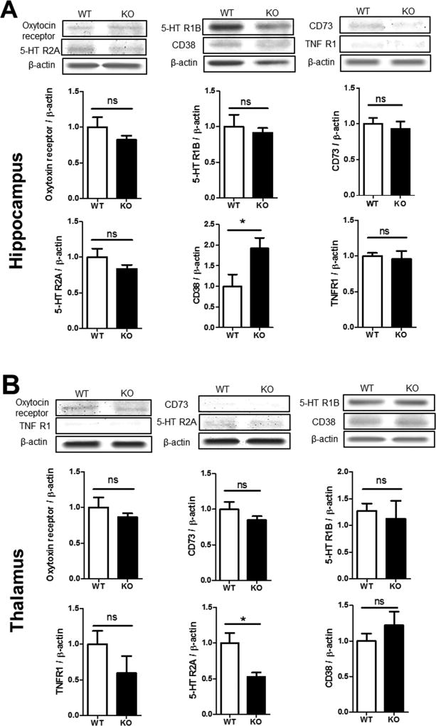
Figure 6. Expression of social behavior genes in the hippocampus and thalamus.
Western blot analysis was performed on brain tissues from the hippocampus and thalamus areas of WT and GluN3A KO mice. A. Western blot images and quantification of the band intensity compared to the basal level. The protein level of oxytocin receptor, serotonin receptors 5-HTR2A, 5-HTR1B, TNFR1 and CD73 showed no statistically difference between WT and KO mice. The level of CD38, however, was significantly higher in the KO hippocampus. N=6, Student t test; * p<0.05, F=1.299. B. The above assay was repeated in the thalamus. All measured protein expressions were similar between WT and KO mice, expect the 5-HTR2A level was significantly lower in this brain region. N=6; Student t test; * p<0.05, F=5.650.

