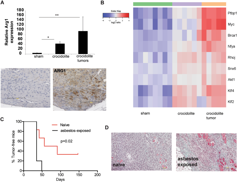
Fig. 6.
Increase in Arg1-positive cells in both inflamed mesothelial and tumor tissues. a Relative expression of Arg1 was verified by q-PCR. Arg1 was highly increased in crocidolite-exposed tissue and expression was maintained in tumors Mean ± SE. N = 5–8 mice. *p < 0.05, **p < 0.01, Mann–Whitney test compared to sham. Arginase 1 (Arg1)-positive cells were detected in the tumors. b Marker genes for self-renewing macrophages [41] shows increased expression in crocidolite-exposed tissues and tumors. c Kaplan–Meier graph of tumor-free mice survival after challenging with RN5 mesothelioma cells (1 × 10⁶) mice exposed to asbestos that had not developed a tumor until 49 weeks after the first exposure to asbestos vs. naïve animals. d Goldner staining of mesothelioma grown in naïve vs. asbestos-exposed mice

