
Fig. 6.
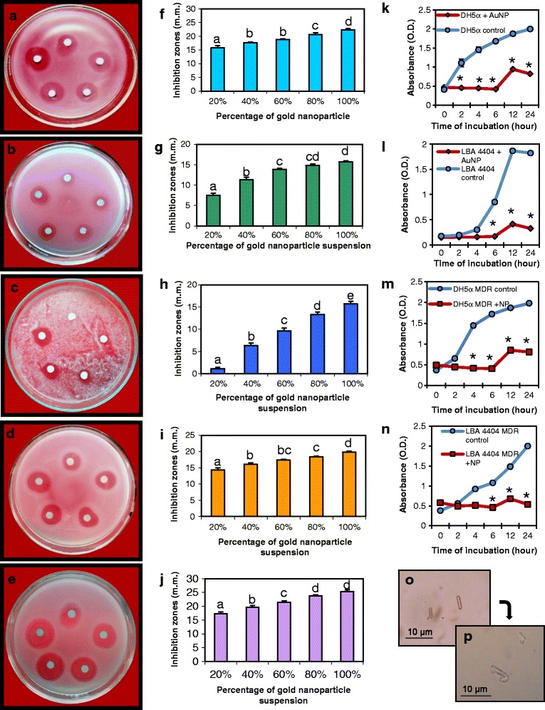
Assay of antimicrobial properties of AuNPs on pathogenic bacteria, fungi, and multi-drug-resistant (MDR) bacteria. a, f Plates and corresponding graphs showing disc-diffusion assay of the nanoparticles with increasing inhibition zones for E. coli. Inhibition zones obtained in similar assays with b, g Agrobacterium tumefaciens. c, h Magnaporthe oryzae. d, i MDR E. coli. e, j MDR A. tumefaciens. All experiments were done with increasing amounts of AuNPs on paper discs; clock-wise from top: 0.249 μg (20%), 0.498 μg (40%), 0.747 μg (60%), 0.996 μg (80%), and 1.245 μg (100%) of AuNPs. Data are means ± SE of three replicates. Different letters indicate statistically significant differences among the samples (P < 0.05, Tukey’s HSD test). Effect of AuNPs on the growth curve of k E. coli, l A. tumefaciens, m MDR E. coli, and n MDR A. tumefaciens. Asterisks indicate significant differences to control (Student’s t test, P < 0.05). o Microscopy of control A. tumefaciens cells. p A. tumefaciens showing loss of cellular integrity after treatment with the AuNPs