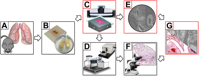
Figure 1.
Workflow of microCT guided sectioning. (A) Lungs were dissected from FVB mice. (B) Specimens were PTA stained, dehydrated and embedded either in paraffin or resin. (C) A microCT scan of each sample was performed and the position of interest defined within the 3D reconstructed phase retrieved data sets. (D) The samples were cut at the predefined ROI using either a standard microtome (paraffin) or cutting by grinding in combination with the laser microtome (resin) (left, TissueSurgeon, LLS ROWIAK LaserLabSolutions, image use permitted by LLS ROWIAK LaserLabSolutions under the Creative Commons Attribution License CC BY 4.0). (E) and (F) A virtual slice and a microscope image of the PTA stained lung tissue slices were produced and (G) the “degree of matching” was analysed using the developed block matching approach.

