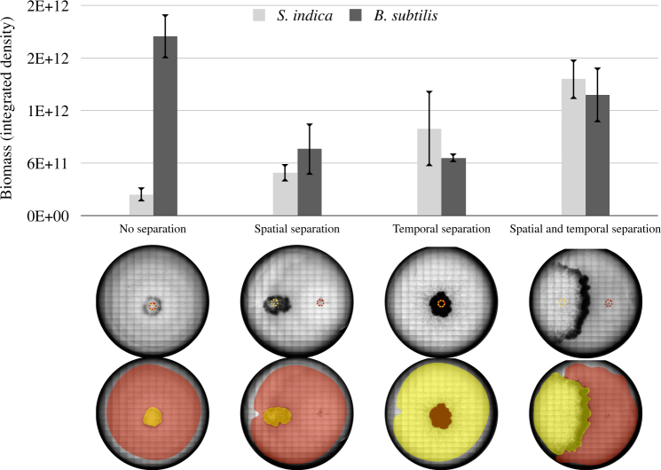
Fig. 6.
Biomass of B. subtilis and S. indica under different spatiotemporal culturing cases. “No separation” refers to B. subtilis culture and S. indica spores being pre-mixed at 1:1 volume ratio, and then inoculated as a single solution. “Spatial separation” refers to approximately 1.5 cm separation of S. indica (left) and B. subtilis (right) inoculation points. “Temporal separation” refers to inoculation of S. indica 3 days prior to B. subtilis inoculation. The yellow dotted circle on the images indicates the S. indica inoculation point. Red dotted circle indicates B. subtilis inoculation point. Below the graph are shown the plate images. The pseudo-coloring indicates S. indica hyphal area (in yellow) and B. subtilis colony (in red). The area of each colonies was manually drawn. Growth of the different species was approximated by tracing their respective colonies on the plate and measuring the image intensity from the engulfing areas 2 weeks after S. indica inoculation. Measurements are from three replicate agar plates, with a representative plate image shown at the bottom. These images show microscopic scans of each plate at 2 weeks of growth. For the “no separation” case, there was no observable S. indica colony expansion after 1 week. We performed four biological repetitions of this experiment, with qualitatively similar results

