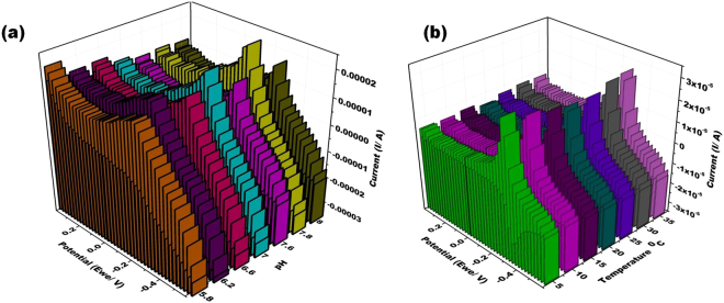
Figure 7.
(a) 3D representation of the cyclic voltammogram at PDNA/MoS2NSs/SPGE for pH of 0.1 M phosphate buffer saline ranging from 5.8 to 8.0 each is having 1 µM MB in the potential range from −0.6 to +0.4 V at the scan rate of 100 mVs−1. (b) 3D representation of the cyclic voltammogram at PDNA/MoS2NSs/SPGE for temperatures ranging from 5 to 35 °C in 0.1 M phosphate buffer saline ranging having 1 µM MB in the potential range from −0.6 to +0.4 V at the scan rate of 100 mVs−1.

