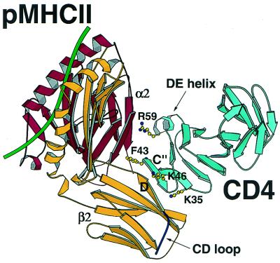
Figure 1.
Ribbon diagram of the CD4–pMHCII complex. The murine I-Ak MHC class II molecule with a CA peptide (green) bound to the antigen-presenting platform interacts with hCD4 (cyan) through both the α2 (red) and the β2 (yellow) domains of pMHCII and domain 1 (D1) of hCD4. Residues Lys-35, Phe-43, Lys-46, and Arg-59 on CD4 D1 essential for binding are highlighted. The CD loop (delineated with an arrow) on the β2 domain of the I-Ak molecule is shown to have no direct interactions with CD4. Note also how CD4 D2 (unlabeled, cyan) makes no contact to pMHCII. All the figures were prepared with molscript (46) and RASTER 3D (47).

