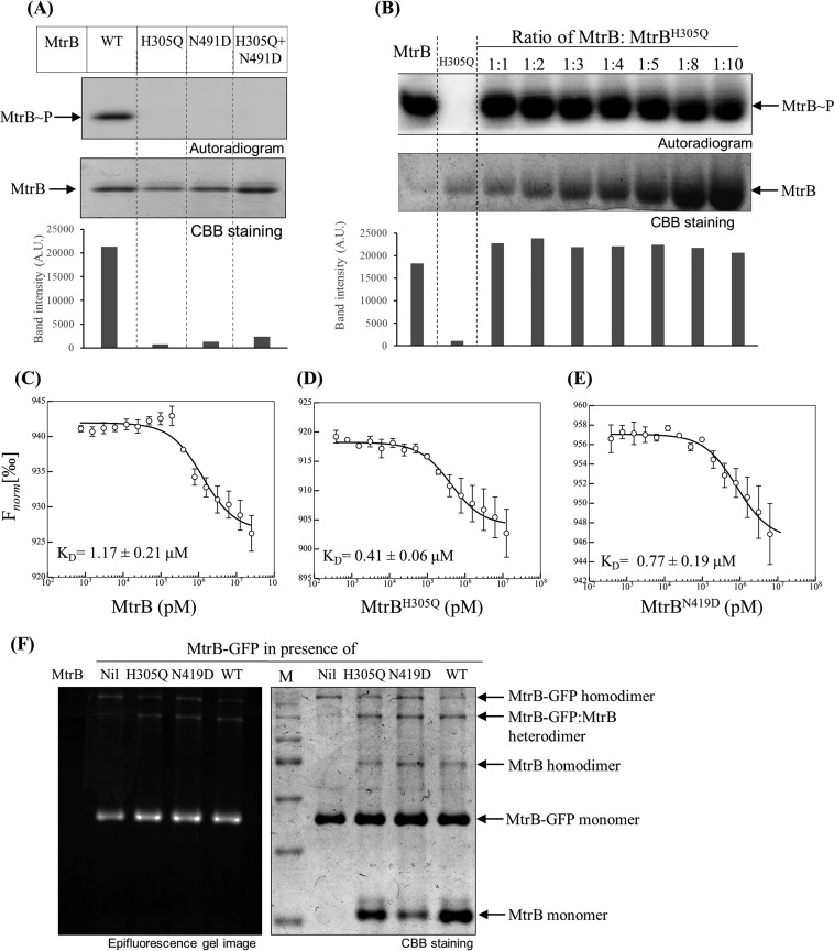
FIG 1 .
Analysis of HK autophosphorylation using mutant complementation assay. (A) Autophosphorylation analysis of MtrB proteins. Wild-type (WT) MtrB or the MtrBH305Q or MtrBN419D mutant was tested in autophosphorylation reaction for 90 min per the protocol in Materials and Methods. The graph below the autoradiogram represents densitometric analysis of the autoradiogram (in arbitrary units [A.U.]). (B) Analysis of trans phosphorylation by competition experiment. MtrB and MtrBH305Q proteins were coincubated in various molar ratios as indicated for 10 min. The autophosphorylation reaction was performed for 60 min. (Top) Autoradiogram; (bottom) CBB-stained gel. The graph below the autoradiogram represents densitometric analysis of the autoradiogram. (C to E) Microscale thermophoresis measurements for determination of interaction affinities of MtrB-GFP with WT MtrB (C), MtrBH305Q (D), and MtrBN419D (E). All graphs were best fit to means ± standard errors of the means (SEM) from three independent experiments. Fnorm, normal fluorescence. (F) MtrB dimerization analysis using nonreducing SDS-PAGE and fluorescence imaging assay by coincubating 2 µM MtrB-GFP with MtrB, MtrBH305Q, or MtrBN419D (at a concentration of 5 µM). Imaging was done per the protocol described in Materials and Methods. Lane M contains molecular size standards.

