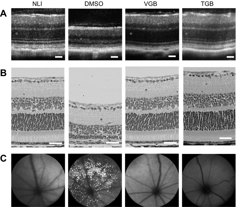
Figure 4.
Preservation of retinal structure in Abca4−/−Rdh8−/− mice pretreated with clinical anticonvulsants before light exposure. A) OCT images of retinal cross-sections. B) H&E staining of retinal tissue slices. Asterisks denote the ONL. C) SLO images of autofluorescent granules in the subretinal space. Images from control animals are repeated from Fig. 2 for clarity. Scale bars, 50 µm.

