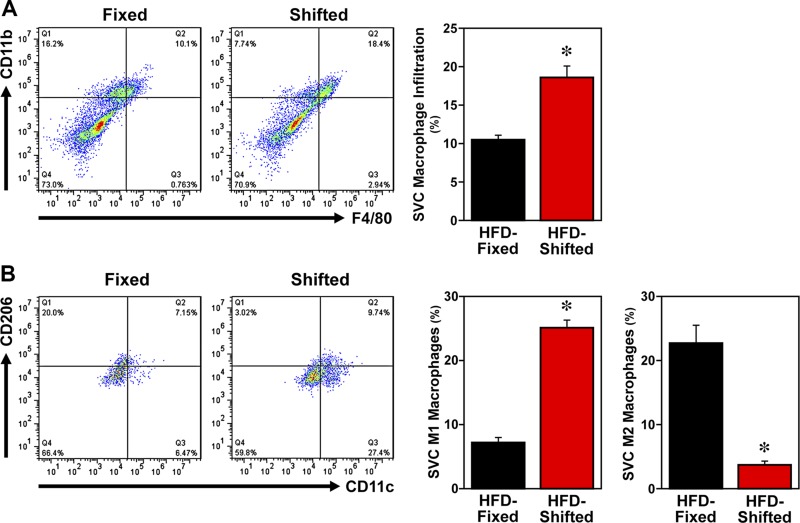
Figure 3.
Effect of shifted LD cycles on adipose tissue macrophage infiltration and polarization in HFD-fed mice. FACS analyses of macrophages in epididymal fat pads from HFD-fed mice exposed to fixed or shifted LD 12:12 cycles. Representative scatter plots of adipose tissue SVCs that were quantified for F4/80 and CD11b expression (A) to identify mature macrophages and for F4/80, CD11b, CD11c, and CD206 expression (B) to differentially analyze proinflammatory (M1) macrophages and anti-inflammatory (M2) macrophages. Bar graphs (right panels) depict quantification of the percentages (means ± sem) of mature macrophages (CD11b+ F4/80+ cells), proinflammatory M1 macrophages (F4/80+ CD11b+ CD11c+ CD206− cells), and anti-inflammatory M2 macrophages (F4/80+ CD11b+ CD11c− CD206+ cells) in adipose tissue SVCs from HFD-fed mice on fixed or shifted LD cycles (n = 3). Asterisks indicate significant differences (P < 0.05) between the fixed and shifted LD groups in SVC macrophage infiltration, M1 macrophages, and M2 macrophages.

