
Figure 1.
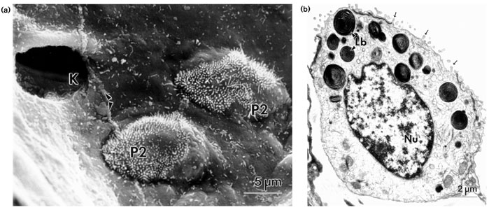
Human lung AE2 cells. (a) Scanning electron micrograph of human lung. Two AE2 cells (P2) are seen to protrude above the largely smooth alveolar epithelial surface. A pore of Kohn (K) and the cell–cell junction (arrowheads) between two AE1 cells are denoted. (b) Transmission electron micrograph of human AE2 cell displaying typical ultrastructural features, such as lamellar bodies (Lb) and apical microvilli (arrows). Nu = nucleus.