Fig. 1.
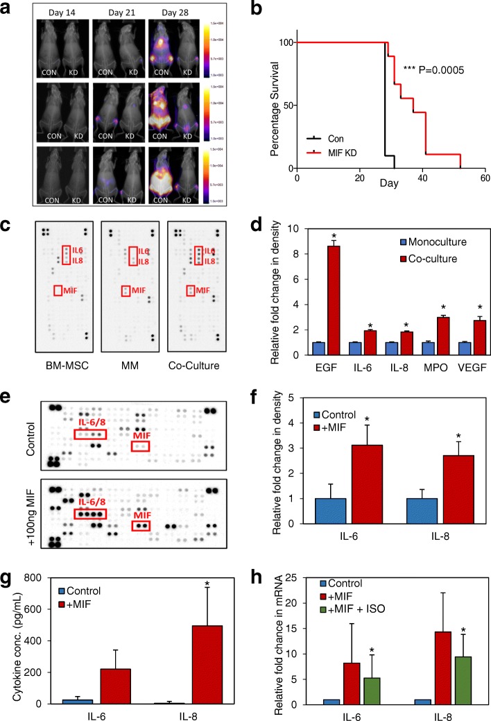
MM derived MIF is pro-tumoral and drives BMSC IL-6 and IL-8. 1 × 10⁶ MM.1S-luc cells (ShE control n = 10, and ShMIF n = 7) were injected via the tail vein of 6–8-week-old NSG mice. a Mice were monitored weekly by bioluminescent imaging. b Kaplan-Meier curve showing survival, analyzed using Mantel Cox regression. c Representative (n = 3) Human XL cytokine array output after a 24-h incubation in either mono or co-culture, cell supernatant was used for analysis. d Graphical representation of c—values for BMSC and MM monoculture intensities were added together and were analyzed against co-culture experiment signal intensity using HL++ image software which show differences in several key cytokines. e, f BMSC were stimulated with 100 ng/mL of human recombinant MIF and incubated for 24 h; supernatant was used for assay. Representative (n = 3) image of cytokine array (e) and subsequent graphical representation (f) of analysis using HL++ software. g Primary BMSC (n = 4) were stimulated with 100 ng/mL recombinant human MIF for 6 h after which IL-6 and IL-8 protein excretion was analyzed by ELISA. h Primary BMSC (n = 4) were incubated with/without 10μg ISO-1 and then stimulated with 100 ng/mL recombinant human MIF for 6 h. IL-6 and IL-8 transcriptional levels were then analyzed by RT-PCR

