Figure 2.
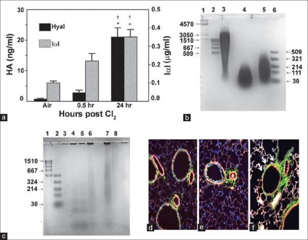
Role of hyaluronan in Cl2-induced lung injury. (a) Detection of HA and IαI, a binding partner of HA, in the BALF of Cl2-exposed mice. Mice were exposed to Cl2 (400 ppm for 30 min) and returned to air. HA was measured in the BALF by ELISA at the indicated times. *,†P < 0.01 compared with air and the value to its left at the same time point, respectively. (b) Agar gel electrophoresis of HA. Lane 1, HA Mega-HA Ladder (Hyalose); lane 2, Select-HA Hi-Ladder; lane 3, HA; lane 4, HA exposed to Cl2 (400 ppm for 30 min) and stored at −4°C for 24 h; lane 5, sonicated HA; lane 6, Select-HA LoLadder, (c) agar gel electrophoresis of concentrated BALF from air and Cl2 exposed mice. Lane 1, Select-HA HiLadder; lane 2, Select-HA LoLadder; lane 3, 95% air-5% CO2 (Air); lanes 4 and 5, immediately post-Cl2; lane 6, 6 h post-Cl2, lane 7, 24 h post-Cl2; lane 8, as in lane 7 but the BALF was treated with hyaluronidase, which degrades HA. In all cases, proteins were visualized with Stains-All (Sigma). (d-f) representative image of mouse airways in naive state (d) or 6 h (e) and 24 h (f) after Cl2 exposure. Increased HA staining (green, arrows) at 24 h in the peribronchial area surrounding airway smooth muscle cells (×200). HA: Hyaluronan; IαI: Inter-α-trypsin-inhibitor; BALF: Bronchoalveolar lavage fluid. Source: Lazrak A, Creighton J, Yu Z, Komarova S, Doran SF, Aggarwal S, et al. Hyaluronan mediates airway hyperresponsiveness in oxidative lung injury. Am J Physiol Lung Cell Mol Physiol 2015;308:L891-903.

