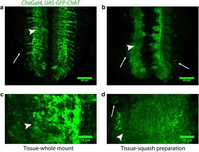
Fig. 1.

Comparison of overall morphology of VNC in whole mount and squash preparation. Confocal images of single optical sections from the VNCs of chaGal4, UAS-GFP-ChAT larvae obtained from whole mount (a, c) and squash (b, d) preparations. a, b Represents VNC at ×1 zoom, while (c, d) represents neuromere hemisegment at ×3.6 zoom. The overall morphology of the tissue and the neuronal connections are retained in the squash preparation. It is judged by scrutinizing the organization of commissures in the neuropil (arrowheads) and cortical region (thin arrows). Magnification ×40 oil objective, N.A. 1.3; Scale bars: a, b 50 µm, c, d 10 µm. The experiments were performed using a set of 5–10 isolated VNCs, and the images represent the majority observation amongst each set
