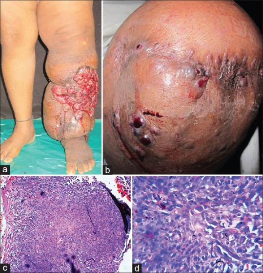
Figure 1.

(a) Elephantiasis of left lower extremity with multiple hemorrhagic nodules on the antero-lateral aspects. (b) Recurrence of multiple hemorrhagic nodulo-ulcerative lesions on the amputation stump. (c) Skin biopsy from the lesion revealed a mass in the deep dermis, consisting of anastomosing blood vessels with endothelial lining showing multiple atypical cells and pleomorphism (×100, H and E). (d) Blood vessels with endothelial lining showing multiple atypical cells and pleomorphism (×400, H and E)
