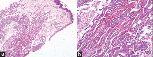
Figure 2.

(a) H and E stain showing proliferation of appendages mainly comprising eccrine glands and capillaries in the mid and deep dermis (×100). (b) H and E stain showing proliferation of eccrine glands, capillaries, and smooth muscles in the dermis (×400)
