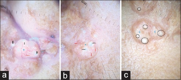
Figure 2.

(a) Dermoscopy of trichoepithelioma shows multiple arborizing vessels (red arrows), milia-like cysts (black arrow) over a whitish background (blue diamond) (polarized, ×10). (b) Multiple branching vessels (red arrow) overlying a whitish background (diamond) with milia-like cysts (black arrow) (polarized, ×10). (c) Multiple milia-like cysts (black circles) overlying a whitish background (star) (nonpolarized ×10)
