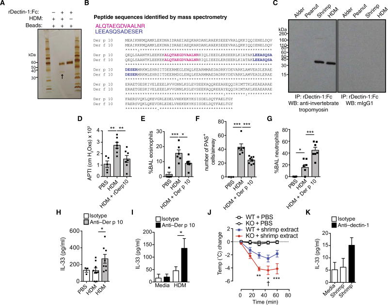
Fig. 5. Identification of invertebrate tropomyosin as a ligand for dectin-1.
(A) Silver stain of a pull-down from rhDectin-1–Fc alone, protein G beads, or rhDectin-1–Fc incubated with HDM extract (see arrow). (B) Peptides identified by MS. (C) Coimmunoprecipitation (IP) of rDectin-1–Fc incubated with alder, peanut, shrimp, and HDM extracts and immunoblotted using anti–invertebrate tropomyosin (anti–Der p 10) or isotype control. WB, Western blot. (D) AHR, (E) BAL eosinophils, (F) lung PAS+ epithelial cells, and (G) BAL neutrophils from male BALB/c mice receiving PBS, HDM (50 µg), or HDM + 10 µg of recombinant Der p 10 intratracheally on days 0 and 5 and harvested on day 7 for analysis. (H) Four hours later, BAL IL-33 levels in male C57BL/6 mice were given PBS or HDM with isotype or anti–Der p 10 antibodies intratracheally. (I) Supernatant IL-33 from 16HBE cells treated for 2 hours with media or HDM in combination with isotype or anti–Der p 10 antibodies. (J) Change in body temperature in Clec7a+/+ (WT) and Clec7a−/− (KO) mice sensitized to shrimp extract. “†” represents death. (K) 16HBE human bronchial epithelial cells treated for 2 hours with PBS or shrimp extract with isotype control or neutralizing anti–hDectin-1 antibodies. Data are representative of two to three independent experiments (A and C) or are means + SEM of two to three independent experiments (I and K) with four replicate wells per condition or pooled from two to three independent experiments (D to G and H and J) and each containing n = 4 to 7 (D to G), n = 3 to 9 (H), or n = 9 to 11 (J) animals per group. *P < 0.05, **P < 0.01, ***P < 0.001, as determined by one- or two-way (I) ANOVA followed by post hoc (Newman-Keuls, Dunnett’s, and Bonferroni) analysis.

