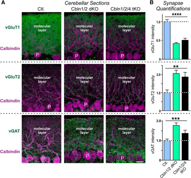
Figure 6.
Double Cbln1/2 and triple Cbln1/2/4 deletions cause a similarly large decrease in vGluT1-staining intensity as a marker of parallel fiber synapses and a similarly large increase in vGluT2 and in vGAT staining intensity as a marker of excitatory climbing fiber and inhibitory interneuron synapses, respectively, in 6-month-old mice. A, Representative immunofluorescence images of sections of the cerebellum from littermate control (left), Cbln1/2 dKO (middle), and Cbln1/2/4 tKO mice (right; age = 6 months). Sections were double labeled for calbindin as a Purkinje cell marker and for vGluT1 (top), vGluT2 (middle), or vGAT (bottom). Scale bar, 20 μm. B, Cerebellin deletions cause opposite effects on the intensity of vGluT1-positive parallel fiber versus vGluT2-positive climbing fiber and vGAT-positive inhibitory synapses. Summary graphs display the staining intensity of vGluT1, vGluT2, or vGAT in cerebellar sections imaged as a function of cerebellin deletions as described in A, All Data are shown as means ± SEM. Synaptic puncta intensity was normalized to the area of calbindin-labeled tissue and to the levels observed in the controls. Statistical analysis was performed by one-way ANOVA (n = 3 mice). **p = 0.0036; ***p = 0.0003; ****p < 0.0001.

