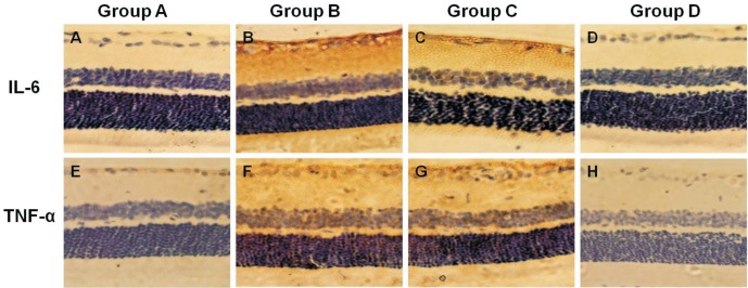
Figure 5. Expression of IL-6/TNF-α in the retina.
A, E: IL-6/TNF-α positive cells were found in the ganglion cell and nerve fiber layers in Group A; B, F: The number of IL-6/TNF-α-positive cells in the ganglion cell layer increased in Group B; C, G; D, H: A marked increase in IL-6/TNF-α-positive cells was observed in Groups C and D; the number decreased especially in Group D compared with Group C. Immunohistochemical staining ×200.

