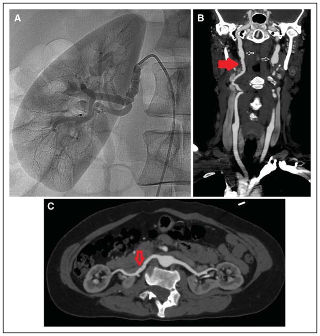
Figure 3. String-of-beads appearance of multifocal fibromuscular dysplasia (FMD).
Selective angiography of the right renal artery (A) and computed tomography angiography of the carotid (B) and renal (C) arteries. B and C are from the same patient. Findings of FMD in the right renal artery are more subtle (C, arrow) but suggest FMD given obvious beading elsewhere (B, red arrow). Also present in the bilateral internal carotid arteries is aneurysmal dilatation (aneurysm vs pseudoaneurysm; B, white arrows).

