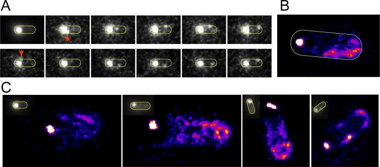
Fig 4. Localization imaging of the nascent new pole.
A) Representative integrated image and filmstrip showing escape of molecules from the brighter old pole and diffusion toward the new pole region. B-C) Localization images of the propensity of Tsr-Venus to reside in the entire cell. The sharp bright dots show an artefactually small localization of the bright stable old pole cluster. At the opposite poles, the areas where the new pole forms show no indication of cluster formational though bulk fluorescence imaging (insets).

