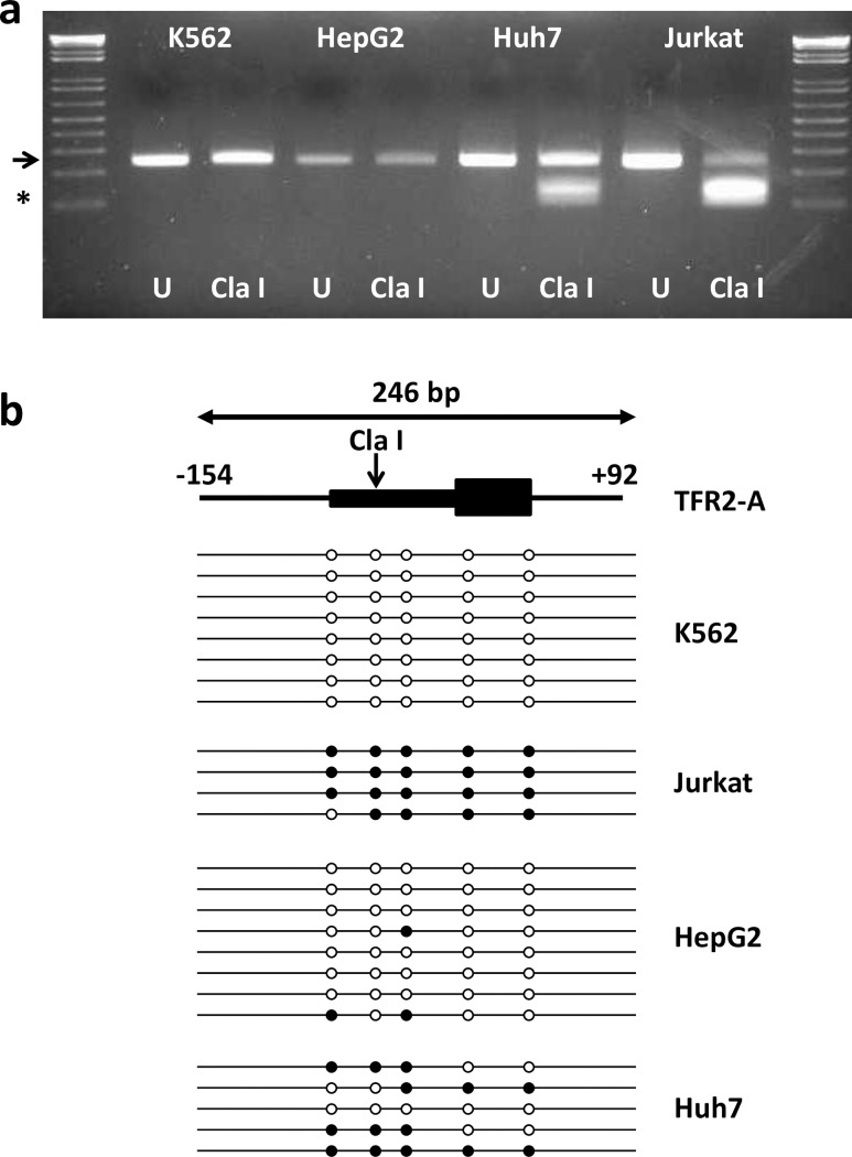
Fig 2. Methylation of TFR2 alpha promoter in human cell lines.
A 246-bp fragment of the human TFR2 alpha promoter was generated by PCR. Amplicons contained a ClaI restriction digest site which will cut the amplicon into 138 & 108 bp fragments if the CpG within the digest site is methylated. Agarose gel (a) shows representative bands from uncut (U; water replacing enzyme) and ClaI-digested bisulphite-converted DNA from K562, HepG2, Huh7 and Jurkat cells. The arrow indicates the position of the full length amplicon (246 bp); * indicates the position of the ClaI digested fragments (138 & 108 bp). The cartoon depicts a representation of TFR2 alpha gene organization and the 246 bp PCR amplicon (b). The 5’ upstream flanking region (from -152 bp relative to the translation start site) and intron 1 (downstream to +92 bp relative to the translation site) are shown as lines; exon 1 containing the promoter and the translated region are shown as boxes. The vertical arrow denotes the ClaI digest site. Horizontal lines below represent bisulphite sequencing of individual amplicons in each cell line. Open circles indicate unmethylated CpGs, filled circles represent methylated CpGs.

