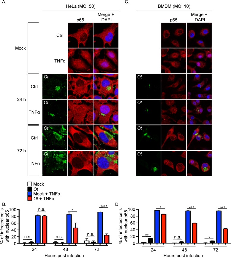
Fig 2. O. tsutsugamushi inhibits TNFα-stimulated p65 nuclear accumulation.
HeLa cells (A and B) or BMDMs (C and D) were infected with O. tsutsugamushi at an MOI of 50 or 10, respectively, or mock infected. At 24, 48, or 72 h, the cells were treated with TNFα or vehicle control (Ctrl) for 30 min after which they were fixed, screened with antibodies against O. tsutsugamushi TSA56 (Ot) and p65, and visualized by confocal microscopy. (A and C) Representative fluorescence images of cells viewed for Ot, p65, and merged images plus DAPI, which stains the nucleus, are presented. (B and D) The mean percentage + SD of cells exhibiting p65 in the nucleus were determined at each time point. Triplicate samples of 100 cells each were counted per time point. Statistically significant (*P < 0.05; **P<0.01; ***P < 0.001; ****P < 0.0001) values are indicated. n.s., not significant. Data are the mean + SD of three independent experiments performed in triplicate.

