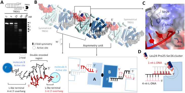
Fig 4. The TREX1-L-structural dsDNA structure.
(A) The nuclease activities of TREX1 in digesting duplex DNAs with 4-nt-long 3′-overhang. (B) The asymmetric unit in the crystal contained 1 TREX1 dimer and 2 ssDNA molecules. The parts shown with a transparent mode depict the symmetry of TREX1 and DNA. The DNA duplex was formed by the ssDNAs bound to 2 TREX1 molecules in separate dimers. The 2 3′-ends of this duplex were 1 nt and 4 nt long and formed an L-like structure at each 3′-terminal, and they are referred to as 1-nt-L-DNA and 4-nt-L-DNA, respectively. (C) The 5′-ends of the duplex region in 1-nt-L-DNA were blocked by Leu24-Pro25-Ser26 cluster. (D) Schematic representation of the 2 modes for TREX1 binding with 1-nt-L-DNA and 4-nt-L-DNA. dsDNA, double-stranded DNA; ssDNA, single-stranded DNA; TREX1, three prime repair exonuclease 1.

