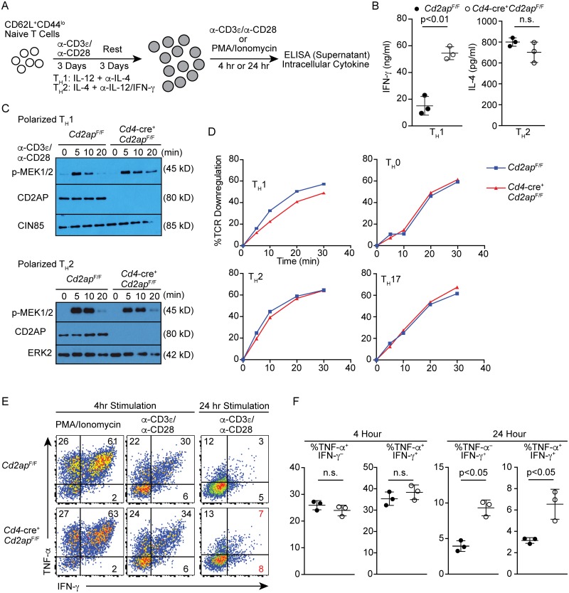
Fig 5. Cd2ap-deficiency causes sustained TCR signaling specifically in TH1 cells in vitro.
(A) Schematic of experimental strategy for culture of CD4 T cells. (B) Concentration of IFN-γ or IL-4 in supernatant measured by ELISA 24 hours after re-stimulation of polarized Cd2apF/F and Cd4-cre+ Cd2apF/F TH1 cells and TH2 cells with plate-bound anti-CD3/anti-CD28 antibodies. (C) Immunoblotting showing phosphorylation of MEK1/2 in polarized Cd2apF/F and Cd4-cre+ Cd2apF/F TH1 or TH2 cells following re-stimulation with plate-bound anti-CD3/anti-CD28 for the indicated times at 37°C. Anti-CD2AP, -CIN85 and ERK were used as control. Data are representative of three experiments. (D) Downregulation of surface TCR of Cd2apF/F and Cd4-cre+ Cd2apF/F T cells that were polarized under indicated conditions. Cells were stimulated with plate-bound anti-CD3 and surface TCR levels were quantitated at the indicated time points. Data are representative of three experiments. (E, F) Intracellular staining for IFN-γ and TNF-α of polarized Cd2apF/F and Cd4-cre+ Cd2apF/F TH1 cells after re-stimulation with PMA and Ionomycin or plate-bound anti-CD3 and anti-CD28 for 4 or 24 hours in the presence of Brefeldin A for the last 2 hours before harvest. Representative plots (E) and statistical analyses with means and standard deviations (F) from three experiments are shown.

