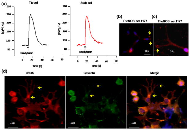
Fig. 9.
Calcium imaging in tip cells: a After 24 h of tip cell formation, EA.hy926 cells were incubated with 10 μM fura 2-AM dye for 30 min, followed by 30 min of washing in fura-free buffer. Images were acquired with the help of ANDOR CCD camera Luca-r attached with Olympus IX71, controlled through Andor IQ software (ANDOR technologies, USA). Bradykinin-induced calcium rise was measured by applying 1 μM of bradykinin. b–d Immunofluorescence imaging of P-eNOS and caveolin. BAECs were allowed to form tip cells. The cells were fixed, permeabilized, and incubated with P-eNOS ser 1177 primary antibody. b Active P-eNOS spots were observed in tip cells compared with that in stalk cells. c P-eNOS expression was found to be dominant at the apex of the tip cell (yellow arrow). d Immunofluorescence for both eNOS (red) and caveolin (green) was done, and the images were captured. Nuclear stain was done using DAPI (blue). The merged image demonstrates colocalization of eNOS and caveolin. (Color figure online)

