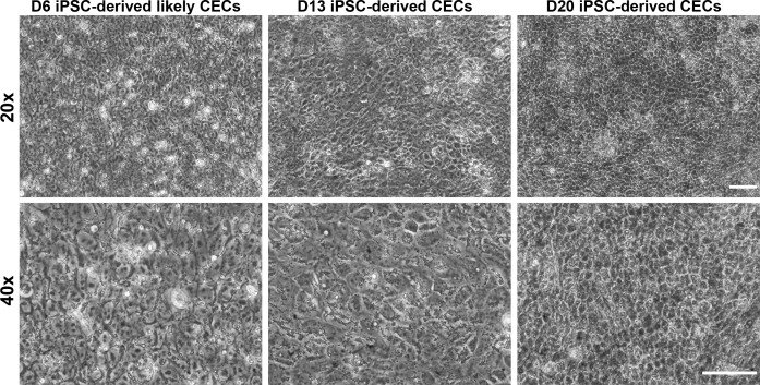
Figure 1.
Generation of CECs from human PBMC-originated, iPSCs. Phase contrast microscopy at various magnifications and time points during CEC differentiation illustrating likely CECs at day 6 (D6) and CECs exhibiting CEC-like hexagonal/polygonal morphology at days 13 (D13) and 20 (D20). Note: The images are of 20× and 40× magnifications and the scale bars represent 50 μm.

