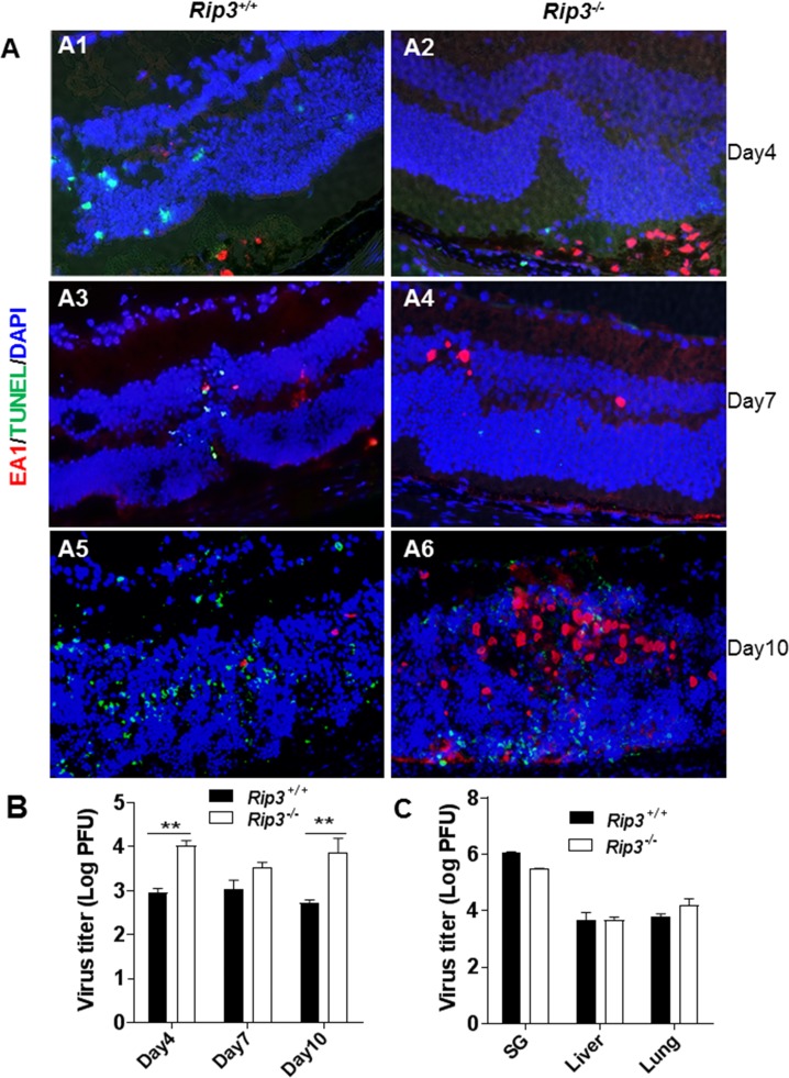
Figure 1.
(A) Merged photomicrographs of staining for MCMV EA (red), TUNEL (green), and DAPI (blue) in MCMV-injected eyes of IS Rip3−/− and Rip3+/+ mice at days 4, 7, and 10 p.i. Fewer TUNEL-stained cells were observed in the inner retina of Rip3−/− (A2, A4) compared to Rip3+/+ eyes (A1, A3) at days 4 and 7 p.i. At day 10 p.i., more infected retinal cells were observed in the injected eyes of Rip3−/− mice (A6) than in the injected eyes of Rip3+/+ mice (A5), and many TUNEL-positive cells were observed in the inner retina of both Rip3−/− and Rip3+/+ eyes. (B) Titer of MCMV (log10 ± SEM PFU/mL) in MCMV-injected eyes of Rip3−/− and Rip3+/+ mice at days 4, 7, and 10 p.i. Data are shown as mean ± SEM (n = 4). Statistical analysis by 2-tailed t-test. **P < 0.01. (C) Titer of MCMV (log10 ± SEM PFU/mL) in salivary glands, livers, and lungs of IS Rip3−/− and Rip3+/+ mice at day 10 p.i. Statistical analysis by 2-tailed t-test indicated no significant difference between Rip3−/− and Rip3+/+ mice.

