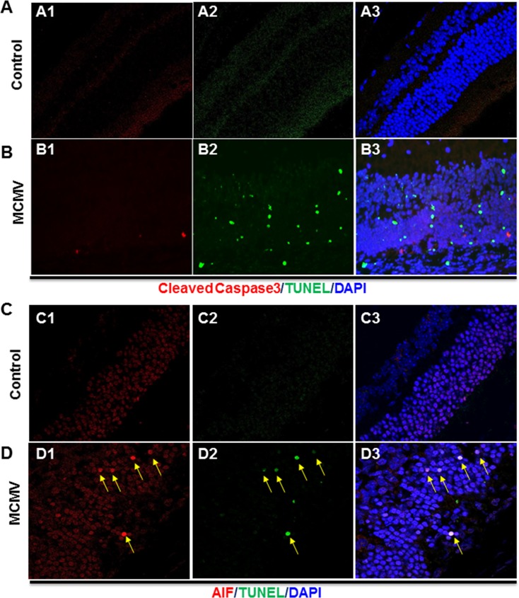
Figure 3.
(A) Photomicrographs of cleaved caspase 3 (A1), TUNEL (A2), and DAPI staining in the mock-injected eye of an IS Rip3+/+ mouse at day 7 p.i. As shown in the merged image (A3), no cleaved caspase 3– or TUNEL-stained cells were observed. (B) Photomicrographs of cleaved caspase 3 (B1), TUNEL (B2), and DAPI staining in the MCMV-injected eye of an IS Rip3+/+ mouse. As shown in the merged image (B3), only a small number of cleaved caspase 3–stained cells were observed in the inner retina and the majority of TUNEL-stained cells did not have staining for cleaved caspase 3. (C) Photomicrographs of AIF (C1), TUNEL (C2), and DAPI staining in the mock-injected eye of an IS Rip3+/+ mouse. As shown in the merged image (C3), no colocalization of AIF and DAPI was observed. (D) Photomicrographs of cleaved caspase 3 (D1), TUNEL (D2), and DAPI staining in the MCMV-injected eye of an IS Rip3+/+ mouse. As shown in the merged image (D3), AIF was colocalized with DAPI in the majority of TUNEL-stained apoptotic cells in the outer nuclear layer (indicated by arrows).

