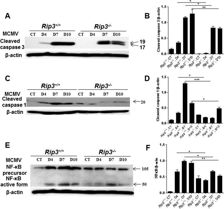
Figure 5.
Western blot stained with antibodies against cleaved caspase 3 (A), cleaved caspase 1 (C), and NF-κB (E) in the injected eyes of mock-injected and MCMV-injected IS Rip3+/+ or Rip3−/− mice at days 4, 7, and 10 p.i. Ratio of cleaved caspase 3 to β-actin (B), cleaved caspase 1 to β-actin (D), and NF-κB to β-actin (F). Data are shown as mean ± SEM (n = 4) and compared by ANOVA. **P < 0.01, *P < 0.05.

