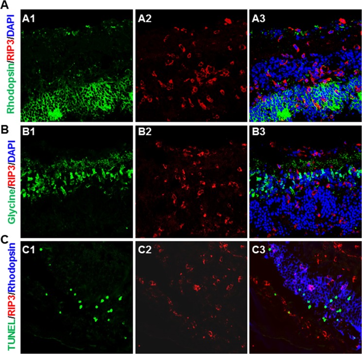
Figure 6.
(A) Photomicrographs of rhodopsin (A1), RIP3 (A2), and DAPI staining in the MCMV-injected eye of an IS Rip3+/+ mouse at day 7 p.i. As shown in the merged image (A3), rhodopsin+ photoreceptors were rarely colocalized with RIP3 staining. (B) Photomicrographs of glycine (B1), RIP3 (B2), and DAPI staining in the MCMV-injected eye of an IS Rip3+/+ mouse at day 7 p.i. As shown in the merged image (B3), glycine-stained amacrine cells were rarely colocalized with RIP3 staining. (C) Photomicrographs of TUNEL (C1), RIP3 (C2), and rhodopsin staining in the MCMV-injected eye of an IS Rip3+/+ mouse at day 7 p.i. As shown in the merged image (C3), TUNEL-stained cells in the outer nuclear layer were not colocalized with RIP3 staining.

