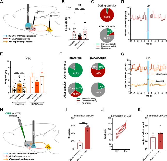
Figure 4.
Activation of D2-MSN terminals in the VP increases motivation. A, Schematic representation of the in vivo single-cell electrophysiological recordings with optogenetic manipulation of NAc D2-MSNs cell bodies. B, NAc D2-MSN optical stimulation (40 Hz, 12.5-ms pulses for 1 s) decrease net firing rate of VP neurons. C, 93.3% of VP cells decrease firing rate and 6.7% did not respond to stimulation (n = 30 cells/four rats). D, Time histogram showing the number of events in the VP before, during, and after a 40-Hz stimulus of NAc D2-MSNs. E, D2-MSN optical stimulation increase the net firing rate of pDAergic neurons of the VTA, with no significant changes in the net firing rate of pGABAergic neurons (npDAergic = 29 cells/four rats; nGABAergic = 5 cells/four rats). F, 82.8% of pDAergic cells increased firing rate in response to stimulation; most of cells returned to baseline activity after the stimulus. pGABAergic neurons presented a majority of inhibitory responses to D2-MSN stimulation. G, Time histogram showing the number of events in the VTA before, during, and after a 40-Hz stimulus of NAc D2-MSNs. H, Strategy used for optogenetic stimulation of D2-MSN terminals in the VP (D2-ChR2 NAc-VP group). I, Optogenetic activation of D2-MSN-VP terminals during cue exposure strongly enhanced breakpoint. J, All animals increase breakpoint in the session with stimulation (ON) versus nonstimulation session (OFF). K, Number of pellets consumed in the PR session was similar between groups. (nD2-eYFP NAc-VP = 6; nD2-ChR2 NAc-VP = 8). Error bars denote SEM; ***p < 0.001 (Extended Data Fig. 4-1).

