
Fig. 2.
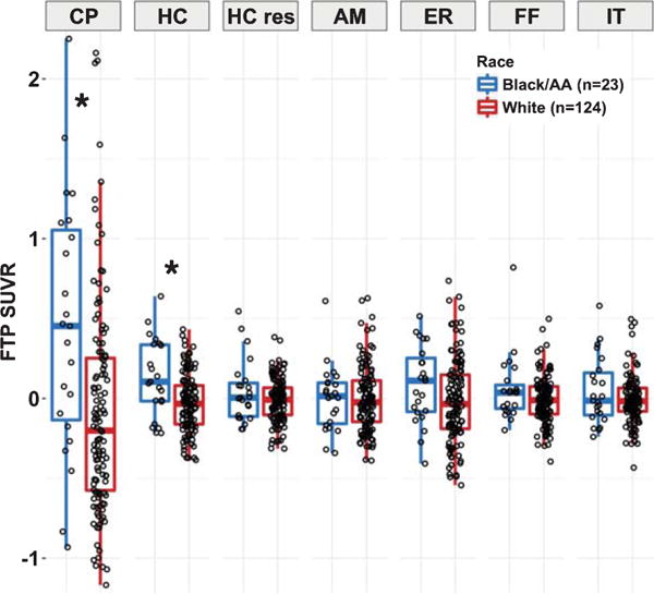
Regional FTP SUVR comparisons between race groups. FTP SUVR (pvc; with PiB DVR, age, sex, and education regressed out) is presented for the Black/AA (n = 23, blue) and White (n = 124, red) race groups. Significant differences (p < 0.05) between races are noted by the asterisks. Elevated FTP measurements are observed in the CP and HC, but not the HC residuals or other ROIs. CP, choroid plexus; HC, hippocampus; HC res, residualized hippocampus; AM, amygdala; ER, entorhinal; FF, fusiform; IT, inferior temporal.