
Figure 3. PAM recognition by WT AsCpf1 and AsCpf1 variants.
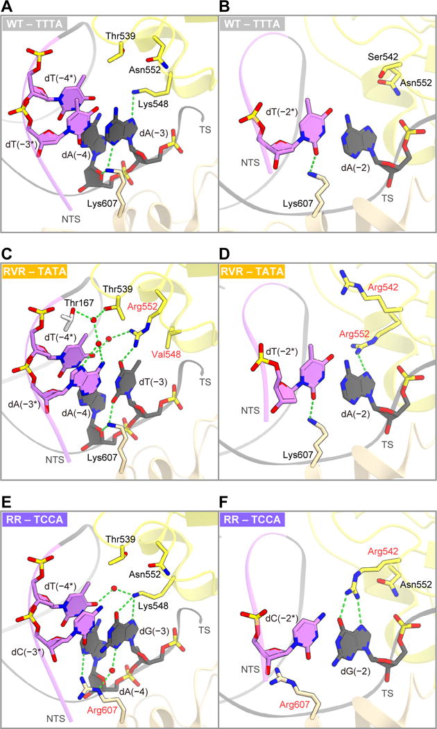
(A and B) TTTA PAM recognition by WT AsCpf1 (Yamano et al., 2016) (PDB: 5B43).
(C and D) TATA PAM recognition by the RVR variant.
(E and F) TCCA PAM recognition by the RR variant.
The interactions with the nucleotides at the first and second PAM positions are shown in (A), (C) and (E). The interactions with the nucleotides at the third PAM position are shown in (B), (D) and (F). In (C)–(F), the substituted residues are highlighted by red labels. In (C) and (E), water molecules are depicted by red spheres.
See also Figure S3.