
Figure 4. Structural differences between WT AsCpf1 and AsCpf1 variants.
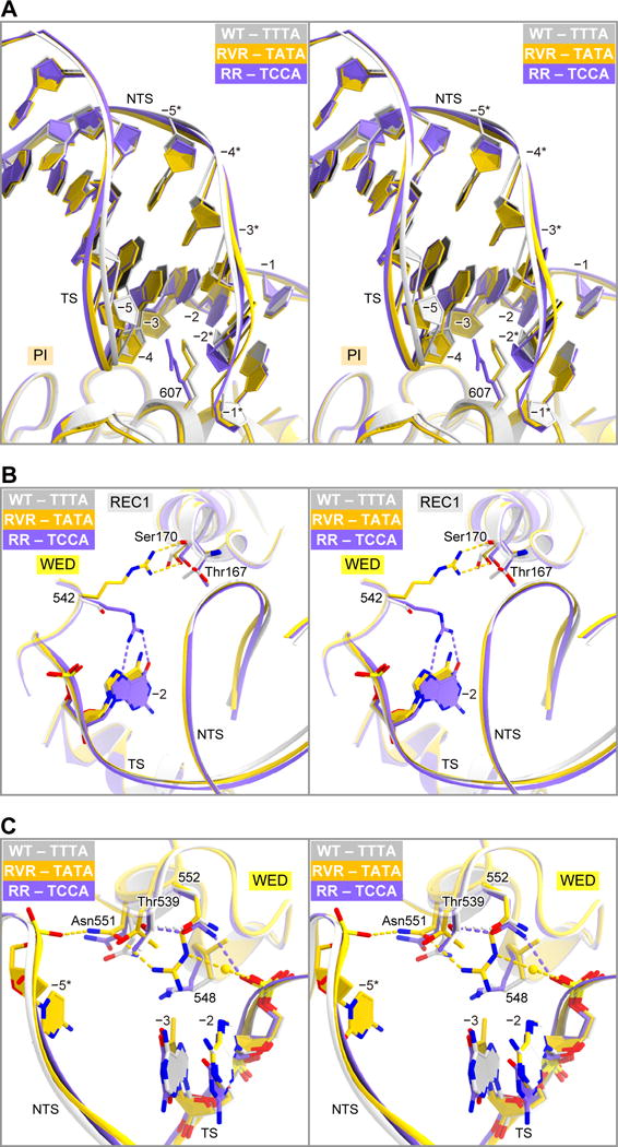
(A) Conformational differences in the PAM duplexes in the structures of WT AsCpf1 (PDB: 5B43) (stereo view).
(B) Structural differences in Arg542 (S542R) between the RVR and RR variants (stereo view).
(C) Structural rearrangements around Arg552 (N552R) in the RVR variant (stereo view). A water molecule is shown as a sphere. In (A)–(C), WT AsCpf1 and the RVR and RR variants are colored gray, orange and purple, respectively.
See also Figure S4.