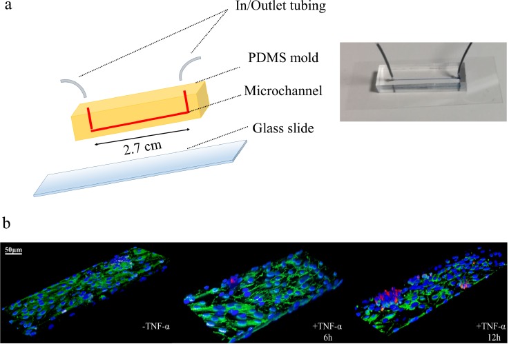
FIG. 1.
Single-channel microfluidic chip. (a) On the left, schematic representation of a single channel microfluidic chip with length l = 2.7 cm, width w = 210 μm, and height h = 42 μm. On the right, a single channel microfluidic chip, with connecting inlet and outlet tubing, filled with a blue ink and placed on the stage of a fluorescent inverted microscope. (b) Representative confocal fluorescent microscopy images of HCT-15 cells (membrane labeled in red with CM-DIL) flowing in the chip and interacting with a confluent layer of HUVECs (nuclei stained in blue with DAPI). VE-cadherin adhesion molecules, arising at boundaries of the endothelial cells, are stained in green [Images are provided for unstimulated (-TNF-α) and TNF-α stimulated HUVECs for 6 (+ TNF-α 6h) and 12 h (+ TNF-α 12h). TNF-α concentration: 25 ng/ml. Scale bar: 50 μm].

