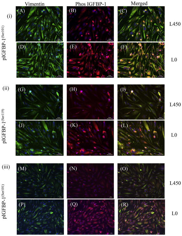
Fig. 10. Dual-immunofluorescence analyses of IGFBP-1 phosphoisoforms in response to leucine deprivation.
Dual immunofluorescence staining depicted the vast majority of decidualized HIESCs to be positive for vimentin (green) also localized IGFBP-1 (red). HIESCs express phosphorylated (i) IGFBP-1(S101) and vimentin (A–F); (ii) IGFBP-1(S119) and vimentin (G–L); (iii) IGFBP-1(S169) and vimentin (M–R). A higher expression of phosphorylated IGFBP-1 at Ser101, 119 and 169 is depicted in decidualized cells in response to leucine deprivation (E, K and Q) compared to decidualized cells cultured in media with leucine (B, H and N). The expression changes in IGFBP-1 were not attributed to the level of decidualization of cells, which was relatively equal in both conditions as shown by vimentin staining (A, D, G, J, M and P). Merged superimposed images (co-localization of red and green yields yellow) are shown in the last panel (C, F, I, L, O and R). Nucleus is stained with DAPI (Blue). Scale Bar = 50 μm. (For interpretation of the references to colour in this figure legend, the reader is referred to the web version of this article.)

